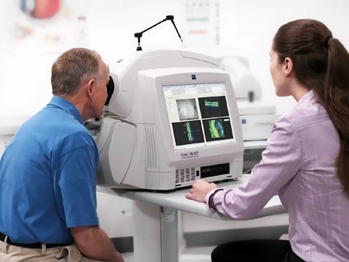
OCT is an eye imaging device that uses a laser light source and has no side effects. It is non-invasive and usually does not require pupil dilation with drops. During the scan, the patient looks at the fixation light inside the device, and the imaging is completed within seconds.
OCT imaging allows us to visualize the deeper layers of the retina that cannot be seen during a standard eye exam. The retinal layers are displayed in three dimensions. Retinal thickness is measured in microns. OCT can detect abnormalities such as edema or bleeding between the retinal layers.
OCT helps in diagnosing diseases such as macular degeneration, central serous retinopathy, diabetic retinopathy, vascular occlusions, vitreoretinal traction, and macular holes. It can also show the stage of these diseases. With OCT angiography, drug-free imaging of the eye’s blood vessels is possible.
OCT is also useful in glaucoma (high eye pressure). It can measure the thickness of the retinal nerve fiber layer, helping to determine the stage of the disease and monitor its progression. OCT measures the cupping at the optic nerve head caused by glaucoma and records the disease’s progression. Additionally, OCT can measure the iridocorneal angle without any contact lens on the eye.
The iridocorneal angle is where the intraocular fluid drains. In eyes with narrow angles, if the angle closes, an acute glaucoma attack can occur. If not treated promptly, the optic nerve can be damaged, leading to vision loss.
OCT can also measure corneal thickness. Recent studies have shown that thinning of the retinal nerve fiber layer can occur before the onset of diseases such as Alzheimer’s and Multiple Sclerosis. Measuring the thickness of the retinal nerve fiber layer can assist in the early diagnosis of suspicious neurological conditions.
Copyright 2026 Private Atagöz Medical Center. All rights reserved.
 Private Atagöz Medical Center